Cuboid Syndrome

Cuboid Syndrome

Cuboid syndrome refers to the dislocation or subluxation of the cuboid bone. The cuboid bone is a cube-shaped bone located on the outer side of the foot. The cuboid is one of the seven tarsal bones which form the hind part of the foot. It is located in front of the heel bone (calcaneus), to which it is joined through the calcaneo-cuboid joint.

Cause:

Cuboid syndrome is a common problem faced by athletes and ballet dancers. It is considered to be a result of inversion ankle sprains (excessive inward twisting of the ankle), which accounts for about 40% of all the injuries affecting athletes.

A brief overview of foot anatomy helps to understand the probable mechanism of cuboid bone dislocation. The peroneus longus is a muscle of the lower leg that runs on the outer (lateral) surface from knee to ankle. At the ankle the tendon of the peroneus longus muscle shifts from vertical to horizontal direction, passing on the side and then below the cuboid bone (at the point of the peroneal groove) and travels obliquely towards the inner (medial) side of the foot to attach to the bone. The cuboid bone acts as a pulley for this tendon. As the muscle contracts the tendon is pulled outward and upward, as a result the tendon rolls the cuboid inwards and downwards. In normal movements, the cuboid is held in place by ligaments.

On sudden inward twisting of the foot, the peroneus longus muscle contracts as a reflex, to reverse the foot to a flat position on the ground. Muscle contraction leads to a strong pull on the tendon, which then throws the cuboid in an inward and downward direction. The ligaments holding the bone in place rupture, leading to a downward displacement or subluxation of the cuboid.

This condition can also result from repeated stress as occurs in ballet dancers, whose ligaments become weak due to excessive and repeated strain from their pointe work.

Symptoms:

  • Dull pain in the centre portion of the outer border of the foot (pain develops suddenly or gradually following an ankle sprain)
  • Pain may radiate to surrounding structures as well.
  • Weakness in the foot especially, as the toes lift off the ground during walking Swelling, or rarely, bruising
  • Standing on toes or rolling the arches of the foot outwards elicits or increases discomfort

Diagnosis:

Cuboid syndrome is often misdiagnosed. X-rays, CT scans or MRI are not much help in diagnosing cuboid syndrome, although they may help to rule out a fracture or tendonitis.

Physical examination may reveal swelling at the site. There is pain on pressing the cuboid bone upwards. Movement of the cuboid is quite restricted (locked cuboid) compared to the other normal foot. In case of severe dislocation, a shallow depression is visible on the upper surface of the foot at the site of the cuboid.

Treatment:

Emergency treatment involves cessation of the activity and finding pain relief through RICE – that is, rest and ice application, applying compression and elevating the foot. Anti-inflammatory drugs also help with pain and swelling.

The dislocated cuboid is manipulated back into position through a procedure called the cuboid whip, however this should only be performed by an experienced clinician.

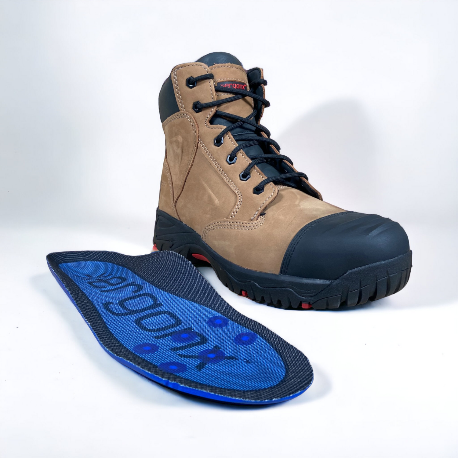
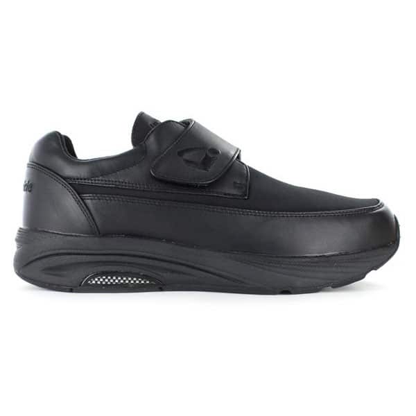
Use of innersoles such as a cuboid pad attached to the orthotic insole, helps to elevate the cuboid to its normal position. Use of innersoles keeps the manipulated cuboid at its place and quality footwear provides sufficient rear foot control.

As with most sports injuries the earlier the condition is treated, the better the outcome.