Anatomy of the Calcaneus
The Calcaneus or heel bone is one of the seven tarsal bones of the foot. Tarsal bones are present between the bones of the leg (tibia and fibula) and the five metatarsal bones, and are collectively referred to as tarsus of the foot.
The Calcaneus is the largest of all the bones in the foot. It is roughly cuboidal in shape and has six distinct surfaces.
The superior surface supports a mass of fatty tissue and anteriorly has three articular facets, the posterior (C below), middle (B below) and anterior artciular surfaces (A below), which articulate with the corresponding facets on the under surface of talus (another tarsal bone present above the calcaneum).

Figure 2 Superior surface of the calcaneus showing anterior (A), middle (M), and posterior (P) facets of the calcaneus, sulcus calcanei (S) that runs between the middle and posterior facets. The canal formed by sulcus calcanei and overlying talus is the sinus tarsi.

A groove extends in front of the posterior articular facet, known as the Sulcus Calcanei.
The middle articular facet is connected to the body through a projecting process called sustentaculum tali. Lateral to it are attached various ligaments along with Extensor digitorum brevis (extensor muscle of the toes).
The inferior surface is uneven and broader from behind with a transverse elevated area called Calcaneal tuberosity. This tuberosity has a small lateral process and a large medial process.

Muscle attachments on Calcaneal Tuberosity
The abductor digiti quinti (the abductor/flexor muscle of fifth toe) originates from the lateral process and the depression between lateral and medial processes while Abductor Hallucis (abductor muscle of the big toe), Flexor digitorum brevis and plantar fascia arise from the medial process.
Anterior to the processes is attached Quadratus plantae (toe flexor).
The lateral surface constricts anteriorly, is flat and has a tubercle in the centre where calcaneo-fibular liganment is attached. Anteriorly there is an elevated ridge called trochlear process or peroneal tubercle separating two grooves one above superior groove and one below inferior groove.
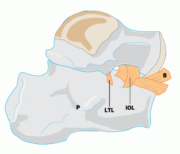
The medial surface is intensely concave to allow for the passage of plantar nerves and vessels to the sole of the foot. Anteriorly on its upper end is a horizontal projection called sustantaculem tali, which from above articulates with talaus and gives attachement to various tendons and ligaments.
Anterior Surface is roughly triangular and articulates with the cuboid bone (a tarsal bone placed in front of calcaneus). The plantar calcaneonavicular ligament is attached on its medial border.
Posterior Surface is very prominent and convex. The lower most part gives attachement to Plantaris muscle whereas in its middle is attached Achilles tendon.

